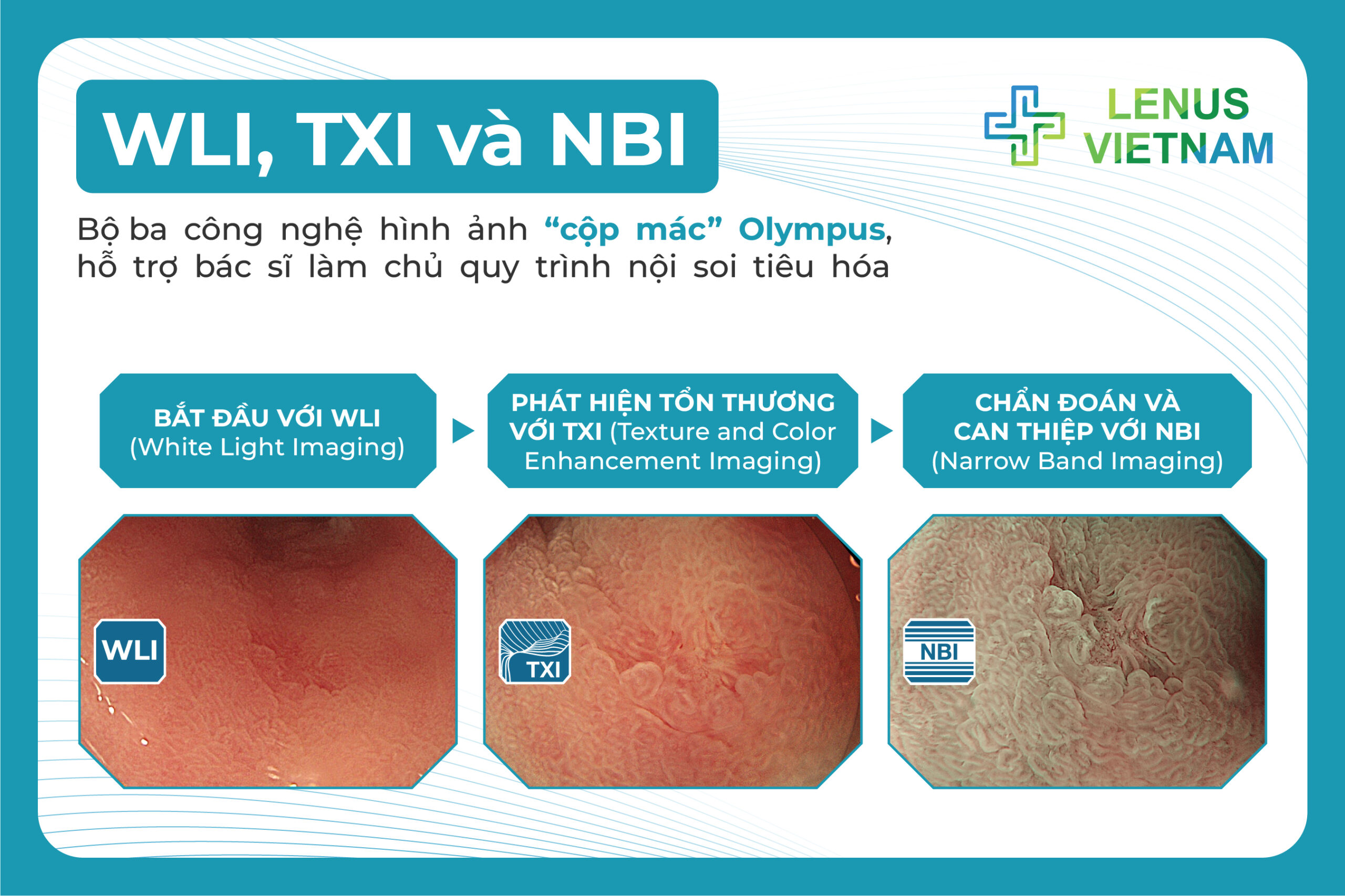
WLI, TXI, NBI: Three key technologies for mastering Endoscopy
With the rapid development of modern endoscopic techniques, gastrointestinal endoscopy today is no longer limited to observing the mucosal surface it has become a comprehensive evaluation process. Physicians can now detect lesions ranging from obvious abnormalities to subtle, flat, and hard-to-recognize changes. Among these advancements, three imaging modes White Light (WLI), TXI, and NBI play crucial roles, supporting physicians from initial screening to early detection, diagnosis, and treatment planning.
White Light (WLI) – the starting point of gastrointestinal endoscopy
Every GI endoscopic procedure begins with WLI (White Light Imaging), which simulates natural light. This is the initial overview step, enabling physicians to assess the structure of the GI tract and identify clear abnormalities such as ulcers, large polyps, or tumors. However, WLI has limitations in detecting small or flat lesions, particularly those blending in with the surrounding normal mucosa.
TXI (Texture and Color Enhancement Imaging) – boosting early detection
When enhanced detection is required, TXI becomes a powerful ally. This technology accentuates surface texture and increases color contrast, helping physicians identify subtle mucosal abnormalities that white light alone may overlook. During early cancer screening, TXI serves as a refined “color magnifier,” maintaining a natural image while improving diagnostic accuracy.
NBI (Narrow Band Imaging) – for diagnosis and guiding intervention
Once a suspicious lesion is identified, physicians switch to NBI. This technology uses specific light wavelengths to highlight superficial blood vessels and mucosal microstructures, enabling better assessment of a lesion’s nature: benign or malignant, invasive or non-invasive.
When combined with magnifying endoscopy, NBI allows physicians to observe even the smallest mucosal changes and to target biopsies with high precision. It also forms the basis of widely used classification systems in early cancer screening, such as JNET, VS Classification, and MESDA-G. These systems support on-the-spot treatment decisions: proceeding with EMR/ESD for resection, or performing biopsy for further confirmation.
Three technologies – one comprehensive endoscopic process
The sequential use of White Light → TXI → NBI provides optimal effectiveness in a GI endoscopy procedure. From detection → highlighting → characterization, this combination helps physicians improve diagnostic accuracy, detect lesions earlier, and intervene in a timely manner—leading to better patient outcomes.
EVIS X1 – a new-generation endoscopy system integrating WLI, TXI, and NBI
Today, all three imaging technologies are integrated into Olympus’ EVIS X1 system. As an advanced endoscopy platform, EVIS X1 is designed to optimize observation, screening, and intervention workflows. With the latest generation of TXI, NBI, and WLI, EVIS X1 not only upgrades imaging quality but also enables physicians to “see the lesion at the right time, in the right place, and in the right way.”
Modern gastrointestinal endoscopy is no longer simply about “what is seen.” More importantly, it is about timely detection, accurate diagnosis, and expanding therapeutic boundaries—contributing to improved clinical outcomes and enhancing patients’ quality of life.