7 Recommendations for Endoscopic Treatment of Early Gastric Cancer & 9 Recommendations for Endoscopic Treatment of Early Esophageal Cancer by VFDE Announced at VGEC 2026
Within the framework of VGEC 2026, Assoc. Prof. Dr. Thai Doan Ky presented the guidelines issued by the Vietnam Federation for Digestive Endoscopy (VFDE) on the management of early upper gastrointestinal cancers, including 7 recommendations for endoscopic treatment of early gastric cancer and 9 recommendations for early esophageal cancer.
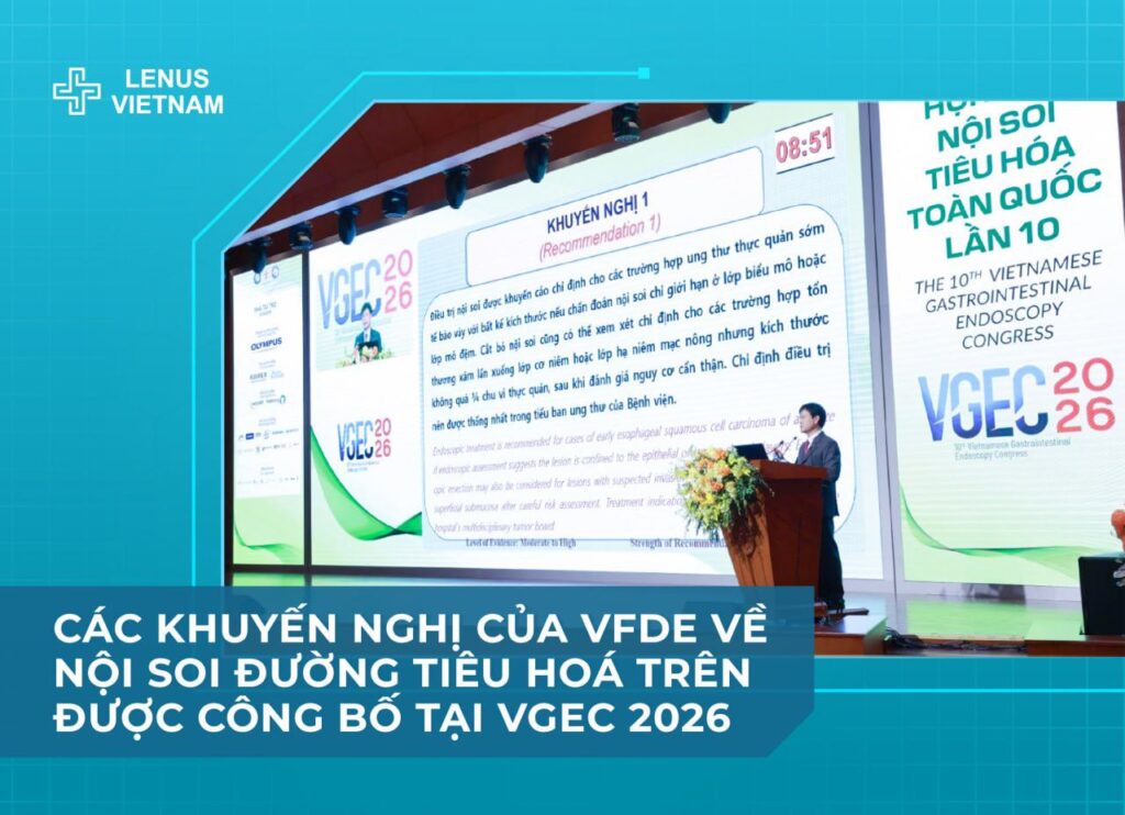
According to Assoc. Prof. Dr. Thai Doan Ky, these recommendations were developed by a panel of physicians and experts through a rigorous process, based on scientific research, evidence, and real-world data both domestically and internationally, with the aim of supporting clinicians in decision-making and optimizing patient outcomes.
Lenus Vietnam recorded these updates from VGEC 2026 and shares them with interested physicians.
7 RECOMMENDATIONS FOR ENDOSCOPIC TREATMENT OF EARLY GASTRIC CANCER
Recommendation 1:
Endoscopic treatment is indicated for differentiated-type early gastric cancer when the lesion is endoscopically assessed as confined to the mucosa (T1a), regardless of size if no ulceration is present, and ≤3 cm if ulceration is present. Endoscopic resection may also be considered for undifferentiated-type early gastric cancer if the lesion is confined to the mucosa, ≤2 cm in size, and without ulceration, provided that histopathological evaluation confirms criteria for curative resection. Treatment indications should be agreed upon by the hospital’s multidisciplinary tumor board.
- Level of evidence: Moderate–high
- Strength of recommendation: Strong
Recommendation 2:
For early gastric cancer, EMR should only be applied to small lesions (≤1 cm) without fibrosis. For lesions >1 cm or with scarring/fibrosis, ESD should be preferred.
- Level of evidence: Moderate–high
- Strength of recommendation: Strong
Recommendation 3:
Chromoendoscopy and image-enhanced endoscopy should be used to assess lesion extent prior to endoscopic resection.
- Level of evidence: Moderate–high
- Strength of recommendation: Conditional
Recommendation 4:
Post-ESD specimens should be processed using a standardized protocol to ensure accurate histopathological assessment. Margin sectioning and reporting should follow the Japanese Classification of Gastric Carcinoma (3rd English edition). Curative resection should be evaluated according to the Japanese eCura classification. Endoscopic resection is considered curative when margins are negative and pathological features indicate no or minimal risk of lymph node metastasis.
- Level of evidence: High
- Strength of recommendation: Strong
Recommendation 5:
Coagulation of vessels at the resection base is required to prevent delayed bleeding. Clip closure should be attempted in cases at high risk of bleeding or perforation. Proton pump inhibitors (PPIs) are indicated to reduce symptoms and prevent complications after EMR or ESD.
- Level of evidence: High
- Strength of recommendation: Strong
Recommendation 6:
For early gastric cancer resected endoscopically, cases with positive vertical margins or high-risk features for lymph node metastasis should be considered for additional surgical gastrectomy. Cases with only positive lateral margins or high-grade dysplasia should be considered for additional endoscopic treatment or close endoscopic surveillance.
- Level of evidence: High
- Strength of recommendation: Strong
Recommendation 7:
Patients who undergo curative endoscopic resection should receive surveillance endoscopy every 6–12 months. Helicobacter pylori infection should be eradicated if present.
- Level of evidence: High
- Strength of recommendation: Strong
9 RECOMMENDATIONS FOR ENDOSCOPIC TREATMENT OF EARLY ESOPHAGEAL CANCER
Recommendation 1:
Endoscopic treatment is recommended for early esophageal squamous cell carcinoma regardless of size if endoscopic assessment indicates confinement to the epithelium or lamina propria. Endoscopic resection may also be considered for lesions invading the muscularis mucosae or superficial submucosa, provided the lesion involves no more than three-quarters of the esophageal circumference and careful risk assessment is performed. Treatment decisions should be made by the hospital’s multidisciplinary tumor board.
- Level of evidence: Moderate–high
- Strength of recommendation: Strong
Recommendation 2:
Endoscopic resection is recommended for high-grade dysplasia of glandular epithelium. In early esophageal adenocarcinoma arising from Barrett’s esophagus, endoscopic resection is indicated when the lesion is predicted to be confined to the mucosa and there is no lymph node metastasis.
- Level of evidence: Moderate–high
- Strength of recommendation: Strong
Recommendation 3:
Image-enhanced endoscopy or Lugol chromoendoscopy is recommended for assessing lesion extent prior to endoscopic resection.
- Level of evidence: Moderate
- Strength of recommendation: Strong
Recommendation 4:
ESD should be the preferred technique for resection of early esophageal cancer with indications for endoscopic treatment. EMR may be an alternative for small lesions ≤10 mm.
- Level of evidence: Moderate
- Strength of recommendation: Strong
Recommendation 5:
The choice of anesthesia or sedation for ESD should be based on multiple factors, including patient condition, lesion location, and size. General anesthesia is recommended to facilitate the procedure and reduce the risk of respiratory complications.
- Level of evidence: Moderate–high
- Strength of recommendation: Strong
Recommendation 6:
Routine use of prophylactic antibiotics and PPIs is not recommended after endoscopic treatment for early esophageal cancer. Their use may be considered in selected patients.
- Level of evidence: Low–moderate
- Strength of recommendation: Strong
Recommendation 7:
Additional treatment is recommended for early esophageal squamous cell carcinoma when histopathology shows submucosal invasion regardless of lymphovascular invasion, or invasion limited to the muscularis mucosae with lymphovascular invasion, even if resection margins are negative.
- Level of evidence: High
- Strength of recommendation: Strong
Recommendation 8:
Endoscopic surveillance is recommended every 3 months during the first year, then every 6–12 months thereafter for patients with curative resection of early esophageal squamous cell carcinoma, to monitor for local recurrence and metachronous lesions, as well as to screen for hypopharyngeal and laryngeal squamous cell carcinoma. CT scan or PET/CT should be added in high-risk cases.
- Level of evidence: Moderate
- Strength of recommendation: Strong
Recommendation 9:
Preventive management of post-ESD strictures and early endoscopic evaluation are recommended for high-risk cases (lesions involving ≥3/4 of the esophageal circumference, length >5 cm, or cervical esophagus location). Local steroid injection is preferred over systemic therapy to minimize adverse effects.
- Level of evidence: High
- Strength of recommendation: Strong